
Paraffin embedding of the whole human cerebral hemisphere to assess arterial distribution territories
DOI:
https://doi.org/10.17179/excli2023-6601Keywords:
tissue processing, whole hemisphere, volume reconstruction, paraffin embedding, brain vascularization, arterial distributionAbstract
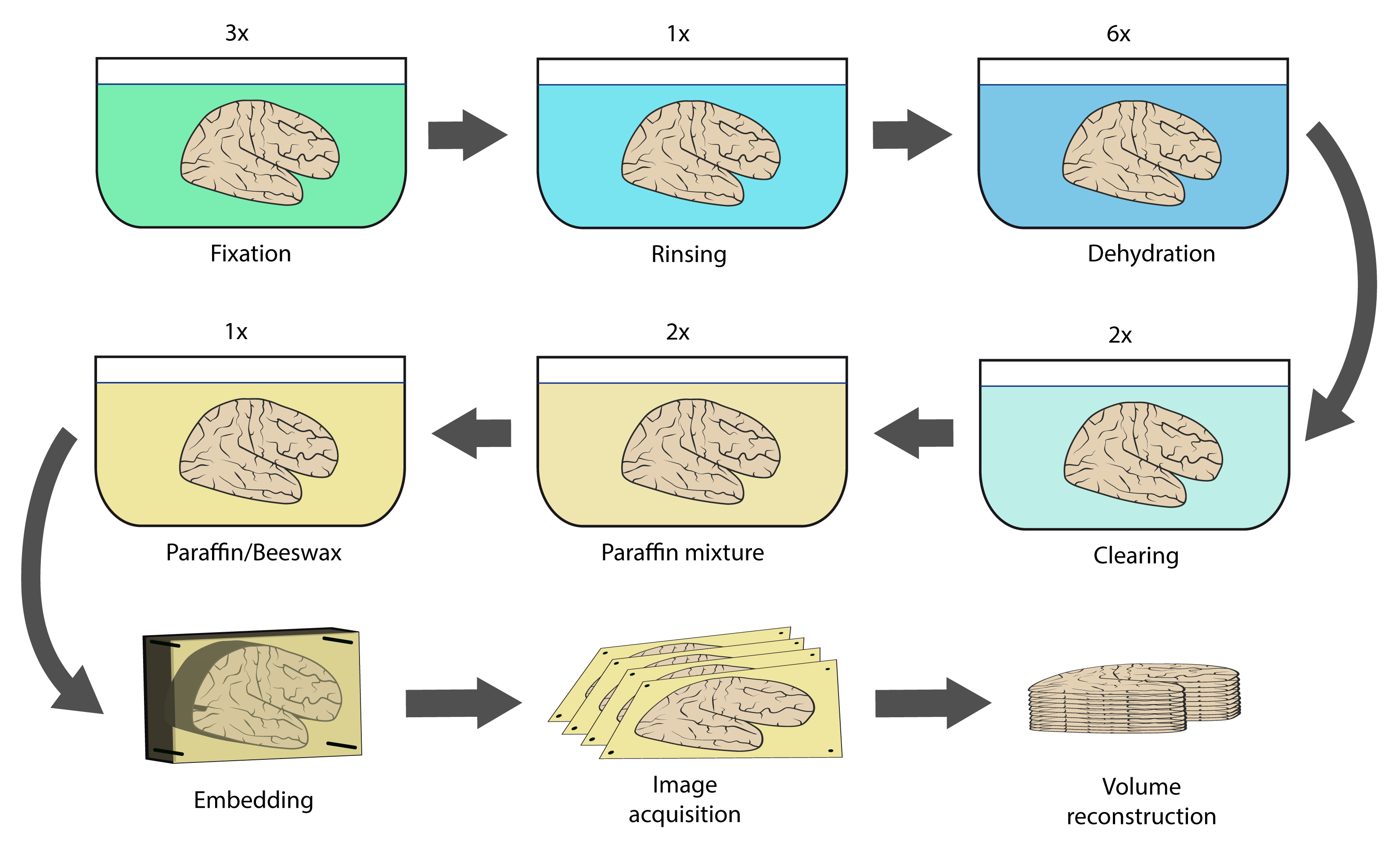
Commonly used to decode the human brain's structural complexity, ex vivo dissection focuses on a given structure or region but cannot depict the whole brain organization (for example, its arterial distribution territories). Where dissection reaches its limit, the combination of tissue sectioning and 3D reconstruction may provide a volume for the assessment of structures from any view angle, following them dynamically to understand their spatial relationships. However, to produce sections, standard histological tissue processing protocols for paraffin embedding cannot be applied to a cerebral hemisphere as the latter is extensively larger than the conventional specimens. This paper presents a protocol for paraffin embedding of the whole human cerebral hemisphere and a method to reconstruct 3D volumes from serially sectioned and photographed paraffin blocks containing embedded hemispheres. Seven ex vivo whole human cerebral hemispheres were included, two were serially sectioned. Main cerebral arteries were injected with colored media to label arterial territories. A detailed description of every step, from tissue processing to image acquisition of cut blockfaces and volume reconstruction, is provided. Tissue processing and section cutting were reproducible, and the former provided complete and homogeneous paraffin wax impregnation. 3D visualization of the reconstructed whole human cerebral hemisphere successfully showed the distribution territories of the main cerebral arteries. In addition, we discuss the challenges we faced and overcame while developing the presented method and highlight its originality.
Downloads
Additional Files
Published
How to Cite
License
Copyright (c) 2024 Mykyta Smirnov, Frédéric Andersson, Laurent Barantin, Igor Lima Maldonado, Christophe Destrieux

This work is licensed under a Creative Commons Attribution 4.0 International License.
Authors who publish in this journal agree to the following terms:
- The authors keep the copyright and grant the journal the right of first publication under the terms of the Creative Commons Attribution license, CC BY 4.0. This licencse permits unrestricted use, distribution and reproduction in any medium, provided that the original work is properly cited.
- The use of general descriptive names, trade names, trademarks, and so forth in this publication, even if not specifically identified, does not imply that these names are not protected by the relevant laws and regulations.
- Because the advice and information in this journal are believed to be true and accurate at the time of publication, neither the authors, the editors, nor the publisher accept any legal responsibility for any errors or omissions presented in the publication. The publisher makes no guarantee, express or implied, with respect to the material contained herein.
- The authors can enter into additional contracts for the non-exclusive distribution of the journal's published version by citing the initial publication in this journal (e.g. publishing in an institutional repository or in a book).


